

What Regenerates First On Planaria Regenerating Animals
Planarian flatworms are well known for their amazing regenerative capacity. In a manner reminiscent of the Wizard'due south Amateur, chopping one worm into piddling pieces volition event in a dish full of tiny worms regenerated from the fragments in simply a few days. In contempo years this system has been rediscovered every bit an experimental model for probing how and why tissues regenerate, with the promise that this volition assistance us to improve tissue repair in our own bodies.
Regeneration in planarians depends on the presence of stem cells called neoblasts. These cells are distributed throughout the body and, when role of the worm has been amputated, they are activated to reform the tissues that have been removed (Wagner et al., 2011). Information technology is still not entirely clear how the stalk cells regenerate specific organs. Are there different types of stalk cell that form different tissues? Do signals produced by nearby cells crusade specific tissues to form? Or is a combination of both stem cell bias and local signalling used? Now, in eLife, Alejandro Sánchez Alvarado and colleagues at the Stowers Institute for Medical Research—including Carolyn Adler as commencement author—have provided new insights into this question past developing a method to specifically remove the pharynx, the feeding organ of the worm, to study organ-specific regeneration (Adler et al., 2014).
The throat itself contains no stalk cells and cannot regenerate the residuum of the worm (Figure 1). However, a worm without a pharynx can chop-chop regenerate this rather circuitous organ. Adler et al. institute that incubating flatworms in sodium azide caused the throat to exist ejected from the trunk without affecting the residue of the worm, thus assuasive them to monitor the process of pharynx regeneration. Blocking stem cell function by irradiation, or past RNA interference knockdown of stem cell-specific genes, prevented regeneration, which suggests that neoblasts have a crucial role in the regeneration of the pharynx.
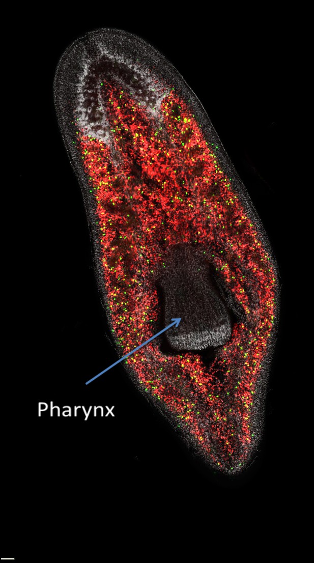
The distribution of neoblasts in a planarian.
Neoblasts are constitute throughout the body simply are excluded from the pharynx. The prototype distinguishes between neoblasts that are in the process of dividing to course new cells (dark-green dots) and those that are not dividing (cherry-red dots) in a freshwater planarian called Schmidtea mediterranea. If the pharynx is removed, the actively dividing neoblasts and their progeny will migrate to the damaged area and begin organ regeneration.
Epitome courtesy of Alex Lin and Bret Pearson.
Local changes in gene expression at the wound site during the early on phases of regeneration revealed that 356 genes were consistently expressed at higher levels than they were under normal weather condition. The importance of each of these genes for regeneration was then tested by employing RNA interference knockdown to inhibit their expression, and using the power of the planarian to feed as a elementary style of measuring how well its throat had regenerated. Twenty genes affected feeding behaviour significantly; some afflicted full general stem jail cell role; other genes affected feeding behaviour but did not touch tissue regeneration; and a small subset of genes specifically affected how the pharynx formed and functioned. The near severe but specific defects in pharynx germination were seen with the knockdown of the gene that encodes a Forkhead transcription factor chosen FoxA.
Forkhead proteins encoded by the FoxA gene family have important roles in specifying the tissues of the digestive system of invertebrates and vertebrates (Kaestner, 2010). In the elementary body of water anemone, FoxA marks the cells that will go on to form the pharynx (Fritzenwanker et al., 2004), while in the roundworm C. elegans, the equivalent of FoxA is key to specifying the dissimilar cell types found in the pharynx during evolution (Horner et al., 1998). In vertebrates, FoxA genes have switched from regulating the pharynx, the entry point to the intestine, to regulating the cells of the intestine itself (Ang et al., 1993).
In mammals, the FoxA family of transcription factors has likewise picked up other functions. These include specifying jail cell fate in the notochord and floorplate, structures that are crucial for centrality formation in the developing embryo (Ang and Rossant, 1994; Weinstein et al., 1994), and controlling the function of adult neurons that produce dopamine (Stott et al., 2013). These divergent functions presumably chronicle to the fact that FoxA proteins are so-called pioneer factors. Pioneer factors bind to DNA at specific sites, opening upward the chromatin structure in many different contexts (Zaret and Carroll, 2011). This ways that they can function in combination with different transcription factors in different tissues. As a pioneer factor, FoxA may have played a fundamental evolutionary office in coordinating the formation of the structures needed to ingest and process food in multicellular organisms, and has so retained and expanded that function throughout evolution.
Planarian FoxA is expressed in the developing and the mature pharynx, and also in scattered neoblast cells around the pharynx that cluster at the site of amputation. In the absence of FoxA, neoblasts are still present, simply they fail to migrate to the amputation site and initiate regeneration, and instead announced to be misdirected to other sites. So is FoxA primarily required for correct cell migration or for correct organ specification? In C. elegans, the equivalent of FoxA (which is called Pha-4) has been shown to bind straight to a broad range of genes that are required for pharynx construction and function (Gaudet and Mango, 2002), suggesting that it has a fundamental part in specifying pharynx prison cell fate. It will be very interesting to explore the direct targets of FoxA in planarians to see if it has a similar master regulatory function. Alternatively, it has been proposed that the evolutionarily conserved function of FoxA lies in regulating particular types of jail cell migration and rearrangement during evolution, rather than primarily in specifying jail cell fate (de-Leon, 2011). Since jail cell behaviour and prison cell fate are closely intertwined, it is hard to separate these functions. However, a more detailed comparing of the targets of FoxA at different stages of planarian regeneration with the corresponding targets in other species could reveal the fundamental roles of this of import transcription gene.
References
Article and author data
Author details
Publication history
- Version of Record published: April xv, 2014 (version i)
Copyright
© 2014, Rossant
This article is distributed under the terms of the Creative Commons Attribution License, which permits unrestricted employ and redistribution provided that the original writer and source are credited.
Metrics
-
- 17,104
- Page views
-
- 361
- Downloads
-
- i
- Citations
Article citation count generated by polling the highest count across the post-obit sources: Crossref, PubMed Fundamental, Scopus.
Download links
A two-role list of links to download the article, or parts of the article, in various formats.
Downloads (link to download the article equally PDF)
Open citations (links to open up the citations from this article in various online reference manager services)
Cite this article (links to download the citations from this commodity in formats uniform with diverse reference director tools)
Planaria: Genes for regeneration
eLife 3:e02517.
https://doi.org/10.7554/eLife.02517
Source: https://elifesciences.org/articles/02517
Posted by: mccluskeyvarty2001.blogspot.com
0 Response to "What Regenerates First On Planaria Regenerating Animals"
Post a Comment